April’s Fungi Focus: Anatomy of a Slime Mould – Trichia decipiens
It is that slime mould time of year again, and so a fitting excuse for me to return to a subject so dear to my heart that I’ve already written about it here and here in previous blog posts, as well as authoring a full-length book on the subject entitled The Creeping Garden to tie in with the documentary film of the same name.
In fact it’s always that slime mould time of year, although different species seem to be more prevalent at different points in the calendar. In March and April, for instance, the False Puffball (Reticularia lycoperdon) is commonly reported. At some point very recently it appears to have inherited the common name of ‘Moon Poo’, derived from the Spanish sobriquet ‘Caca de luna’ under which it is known in certain Mexican communities. Its silvery grey blobs, up to 10cm in diameter and with a slightly pinkish sheen, appear around eye level on standing tree trunks, making it particularly conspicuous at a time when other larger fungi have all but disappeared. The vivid sulphurous yellow Flowers of Tan slime mould (Fuligo septica) is more a Summer species. It too seems to have captured the imagination of a certain sector of British nature lovers, who have come to refer to it as the Dogs Vomit slime, although historically this rather unpleasant label has referred in Britain to a wholly different white species usually found on grass, Mucilago crustacea, with its new application making its way over from the North American vernacular.
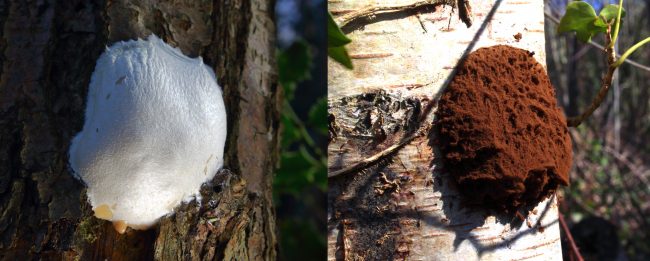
The large fruit bodies of False Puffball, now colloquially referred to as ‘Moon Poo’, before and after releasing spores.
Slime moulds, or myxomycetes, one must emphasise, are not fungi, but they are most commonly found, photographed and recorded by fungi fanatics, primarily because they appear in the same damp wooded spaces where you’d expect to find fungi, even at the times of year when there are few fungi to be found. The key difference is that they are single celled, at least for large parts of their life cycle, and can be recognised by the fact that their fruitbodies grow on their substrate, rather than from it. They reproduce by spores, but that’s effectively where their kinship with members of the fungi kingdom ends. The spores hatch into microscopically small tailed swarmer cells that forage individually through the leaf litter and other decaying organic material, feeding on bacteria, yeasts, fungi spores and other by-products of decay invisible to the naked eye. At a certain point, these individual cells aggregate to form a visible and intricately patterned slimy mass known as the plasmodium that crawls at an almost imperceptible speed as it continues its foraging process. The plasmodium can be a variety of colours such as pink, orange or yellow, but more often than not is white, and in any measure, can seldom be used for species recognition in its own right. For this one has to wait until this plasmodial stage metamorphizes to form the fruit bodies, which really provide slime moulds’ only means of visual identification.
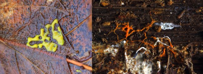
Slime mould plasmodia come in a variety of colours but are never useful alone in identifying a particular species.
We can get an idea that slime moulds must be a lot more common in the natural world than the rare sightings of these fruitbodies would suggest, because I’ve often come across their stray spores on many occasions while looking under the microscope at other fungi samples. Put a pile of damp leaves, twigs and soil in an aquarium, keep moist and leave for a while, and a slime mould of some type or other will probably materialise at some point. Indeed, some people even keep them as pets in this way! They must be everywhere, but the ones that are more commonly reported are the relatively select few with larger fruitbodies over a couple of centimetres in size – the False Puffballs, the Flowers of Tan, the Corals and the Wolfs Milk slimes. The vast majority produce sporangia that are much smaller, really pushing the limits of the macro lens for those attempting to photograph them. Just to give an indication of their myriad shapes, sizes and colours, I’ll direct you to this wonderful website, The Hidden Forest, which includes photos of a number of species found and photographed in New Zealand, although many of the same types crop up all over the world.
In previous posts, I’ve talked about the behaviour of the mobile plasmodium, but now I’d like to turn my attention to the often ingenious ways that they disperse their spores through their fruitbodies. Steven L. Stephenson’s handy introductory book on the subject, Myxomycetes: A Handbook of Slime Molds (1994), differentiates a number of different types of fruit bodies. The aforementioned species like the False Puffball form aethalia when the plasmodium masses together, “a comparatively large fruiting body that is usually hemispherical or cushion-shaped, always sessile, and often rather variable in size.” These typically are covered by a skin beneath which the spore mass forms, with the spores released to be carried by the wind as the fruit bodies dry out and the skin ruptures.
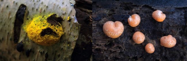
Flowers of Tan and Wolfs Milk slime moulds are two species that form large fruiting bodies called aethalia.
The much smaller more typical species form sporangia however, groups or clusters of “relatively small fruiting bodies of fairly uniform shape and size for a particular species [that] are formed when a plasmodium breaks up into a number of small portions… Depending on the species, sporangia may be sessile or stalked, and a wide range of different shapes and colors are represented by the various myxomycetes that produce this type of fruiting body.”
Identifying species from the tiny sporangia is a challenge, to say the least, given that near on 900 species of slime moulds have been categorised, and that most are very very small, at best about a millimetre or two in height. It really requires a microscope when you’re starting out, although with the myxomycetes you can usually get away with one that is less powerful than one might use for hardcore examination of fungi. (I apologise for my photographic limitations here, but to capture them in their full beauty, as people like the nature photographer Alison Pollack do, you need a powerful macro lens, great light, endless patience, and the use of digital stacking techniques to ensure a good enough depth of field so that you can see all the details).
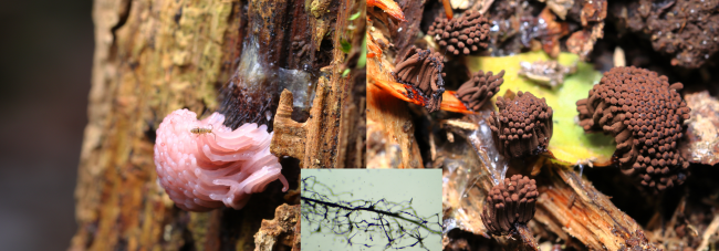
The clustered sporangia fruiting bodies of Stemonitis species turn from pink to brown as the spore mass develops within the mesh arrangement formed by the capillitium, seen under a microscope in the inset photo.
Let us look at a relatively common example, which is the slime moulds falling within the Stemonitis genus. The spore releasing part of the sporangia grows on a stalk. The part of the stalk that grows into the area where the spores are contained is called the columella. With Stemonitis, both stalk and columella are relatively long, with the slender sporangia reaching up to 20mm in height and looking a bit like a feather duster held on stalks as wiry and dark as the hairs in a horse’s mane or tail.
From the columella, a mesh-like cage of branching “threadlike elements” emerge called the capillitium, within which the spores are formed. The sporangia starts out a pinkish colour, but as the spores form, they transform it into a chocolatey brown colour – hence a common name for these types is the Chocolate Tube slime mould. Basically all it takes is a gust of wind for this genus to blow the spores from its mesh of capillitium and thus a new generation of Stemonitis is born. There are a number of separate species in the Stemonitis genus, and you’ll need to look closely at things like relative spore sizes, the diameter of the various threads in the capillitium and other microscopic features to really zero in on whether you have S. fusca, S. axifera, S. splendens or whatever, but let’s not go there for the moment.

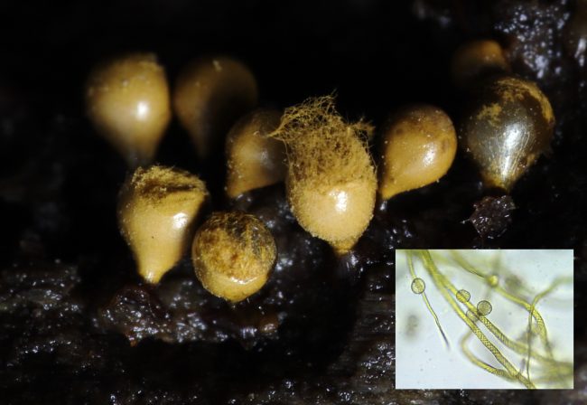
The grainy, slightly shiny look to the minute sporangia of Didymium squamulosum is due to minute calcium crystals on the peridium, seen microscopic detail in the inset photo of the spores of this species.
Another key feature to slime mould identification is the peridium – the skin that encompasses the spore mass. The literature refers to the peridium of the aforementioned Stemonitis as “evanescent”: that is, it vanishes as the sporangia develops to the point that it is really no longer present by the time they’ve turned into their Chocolate Tube shape. In certain species like Didymium squamulosum, the peridium is thin and nearly transparent, but is covered in tiny calcium carbonate crystals, another feature that helps you down the right track to identification. I’m not sure what purpose these crystals serve – possible just to prevent the sporangia from drying out – but they do give the fruit bodies of such species a grainy, slightly iridescent look that is perceptible even without a microscope.
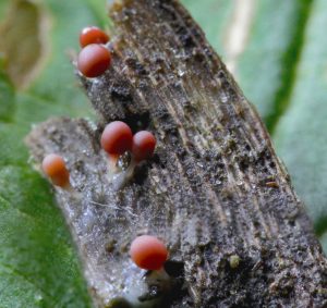
I’ll end with another very common species in the UK, Trichia decipiens, which grows initially as tiny white pear-shaped balloons that turn pink, then yellow to olivaceous brown, as the peridium thickens. The spore mass within it grows, clustered among the capillitium, which in the case of this species are not mesh-like or branching, but consists of a number of single threads - known as elaters - that taper at either end and are covered in spiral bands. The capillitium and spores grow until reaching critical mass, upon which point their force causes the peridium containing them to rupture, and the change in pressure results in the elastic and grooved elaters catapulting the spores out into the atmosphere in a remarkable feat of natural engineering.
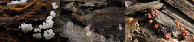
The sporangia of Trichia decipiens developing from white through pink to brown
I should mention that Trichia decipiens should be fairly easy to identify due to its shape, except that its colour is quite variable and that colour alone is not a particularly reliable feature when identifying slime moulds. In fact, there is another type that is considered by some a variant, referred to as Trichia decipiens var. olivacea, and by others as a separate species in its own right called Trichia crateriformis. The key difference is that the peridium ruptures very cleanly around the diameter of the sporangia, as if one were lopping the top of a boiled egg off – a fairly exacting means of differentiating the two species that slime mould-ologists refer to as “circumsessile dehiscence”. One is left with a wine-glass like shape with a candy-floss like arrangement of the mass of elaters covered with the yellow spores that is easy to see under a hand lens.
Myxomycetes identification is a tricky business, and very few are party to its secrets. Hopefully this post will give a few pointers as to where to begin, but there are a variety of good social media groups with some very generous experts giving their help and advice with identification, and some really great books and websites containing photos that reveal the astonishing colours of some of these species. Slime moulds may not be at the forefront of everyone’s mind, but provide a fascinating gateway into appreciating the hidden world of biological activity occurring around us on a daily basis.
Sporangia of Trichia decipiens releasing spores. The “circumsessile dehiscence” indicates this is Trichia decipiens var. olivacea, while the inset photo shows the spores alongside the elaters in microscopic detail.
Comments are closed for this post.
Discussion
How wonderful you’ve found this article Alison. Your photography is fantastic! Mine pales into comparison with such tiny subjects, I’m afraid. I’ve not got the patience for stacking photos, alas.
I’ve heard great things about Secretive Slime Moulds: Myxomycetes of Australia, but yet to get a hold of it. Thanks for the recommendation!
Hi Jasper, This is a fun article on my favorite subject! Thanks very much for the shoutout on my slime mold photography. I am completely smitten and fascinated by myxos and I love to show their incredible beauty, in such a tiny package.
I’ll add one book to your list above, a new one for Australia, but since so many myxos are cosmopolitan this would be useful for other areas as well: Secretive Slime Moulds: Myxomycetes of Australia. It is available now in Australia but not yet in the US where I live, but my Australian myxo friends really like it.
Another very good place to learn about myxos is the excellent Facebook group Slime Mold Identification and Appreciation. There are world experts in there who regularly comment and give advice to myxo fans.
And here is a website where you can see myxos in incredible detail: Myxomycetes.net
Thanks for the kind words about the post Ian. In answer to your question about good entry level spotters guides, alas, there’s not a lot out there. Many fungi guides cover slime moulds in passing – for example, the wonderful Fungi of Temperate Europe has about ten pages on them. Many of the more affordable books devoted to slime moulds only include about a couple of dozen of the near 900 species out there, which makes getting exact identifications for a layman such as myself a frustrating business.
More exacting slime mould books for a specialist market are either out of print, incredibly expensive and/or in a foreign language – for example, what is often described as the best book is a French one, the 2-volume ‘Les Myxomycètes’ : https://www.nhbs.com/les-myxomycetes-2-volume-set-book
Steven L. Stephenson’s Myxomycetes: A Handbook of Slime Molds (1994) is one of the introductory books on the subject, although it contains plain line illustrations rather than photos: https://www.amazon.co.uk/Myxomycetes-Handbook-Stephenson-Stempen-Paperback/dp/B011SIY1IY
My one book The Creeping Garden is more generally about what slimes mould actually are and what kind of research they are being used for, but it is certainly affordable: https://www.amazon.co.uk/Creeping-Garden-Jasper-Sharp/dp/1903254787/
I’ve not seen it myself, but this looks interesting The Curious Observer’s Guide to Slime Mold of UC Santa Cruz and Beyond – remember the same species that can be found in Santa Cruz are largely the same as those found in the UK :https://www.amazon.co.uk/Curious-Observers-Guide-Slime-Beyond/dp/1725169428/ref=pd_sbs_2
Angele Mele’s self-published A Companion to Cosmopolitan Slime Molds is really nice, featuring watercolours by this same artist whose illustrations appeared during the end credits of the Creeping Garden film. It can be ordered here: http://www.sporangela.com/slimemoldprints
And finally Where the Slime Mould Creeps by Sarah Lloyd is another great book with some beautiful photos of the most commonly found species. https://www.nhbs.com/where-the-slime-mould-creeps-book
Hope this all helps!
Jasper
Great article (as ever), thanks. Are you aware of any decent field guide to slime moulds? As a complete beginner anxious to know when/if i’ve seen a slime mould, i’ve been looking for a book for a while…no joy yet…
We have these growing on our shed.
Very strange to say the least as we live in a residential urban area in East Anglia England.
Dawn Kelly
1 May, 2023